Pulmonary Vessel Segmentation based on Orthogonal Fused U-Net++ of Chest CT Images
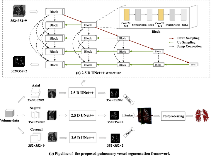 Overall architecture of the proposed pulmonary vessel segmentation framework. Each volume data is sampled as adjacent slice groups along three orthogonal axes andthen fed to a 2.5D U-Net++ network; The features extracted by the three parallel 2.5D networks are fused to optimize the volumetric representation; In the post processing, a structure graph is extracted to refine the segmentation result. (a) The structure of 2.5D U-Net++, whose output is the two-channel probability map of the center slice. (b) The whole framework, including the orthogonal fusion of multi-planar networks and the post-processing for segmentation refinement.
Pulmonary vessel segmentation is important for clinical diagnosis of pulmonary diseases, while is also challenging due to the complicated structure. In this work, we present an effective framework and refinement process of pulmonary vessel segmentation from chest computed tomographic (CT) images. The key to our approach is a 2.5D segmentation network applied from three orthogonal axes, which presents a robust and fully automated pulmonary vessel segmentation result with lower network complexity and memory usage compared to 3D networks. The slice radius is introduced to convolve the adjacent information of the center slice and the multi-planar fusion optimizes the presentation of intra and inter slice features. Besides, the tree-like structure of pulmonary vessel is extracted in the post-processing process, which is used for segmentation refining and pruning. In the evaluation experiments, three fusion methods are tested and the most promising one is compared with the state-of-the-art 2D and 3D structures on 300 cases of lung images randomly selected from LIDC dataset. Our method outperforms other network structures by a large margin and achieves by far the highest average DICE score of 0.9272 and a precision of 0.9310, as per our knowledge from the pulmonary vessel segmentation models available in literature.